
Pacemaker patient assessment with CHART
October 20, 2022
Situation
Female patient with a previous history of carcinoma and mildly decreased systolic function due to cardiotoxicity. She had a previous heart trauma and a dual chamber pacemaker implanted for sick sinus syndrome. Back for control, where a Cardio-HART (CHART) test is performed before Transthoracic Echocardiogram.
CHART report interpretation
Risk Assessment
The patient is 74 years old, hypertensive, dyslipidemic, a former smoker, and under high levels of stress. Medications: Candesartan (16mg), Indapamide (1.5mg), Triflusal (300mg), Atorvastatin (20mg)
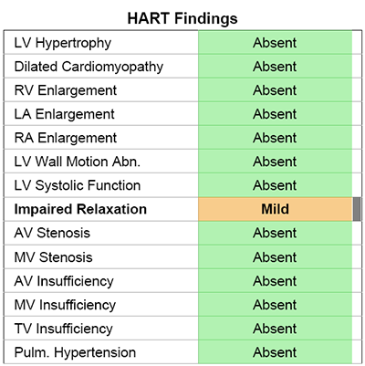
HART-findings and HF prediction
HART-findings shows mild diastolic dysfunction, type impaired relaxation.
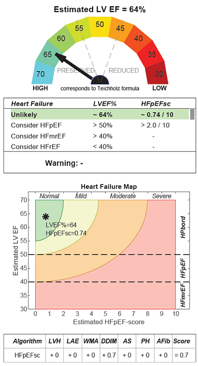
The estimated LVEF shows normal, preserved systolic function (LVEF=64%).
The probability of HFpEF is unlikely according to the CHART assessment.
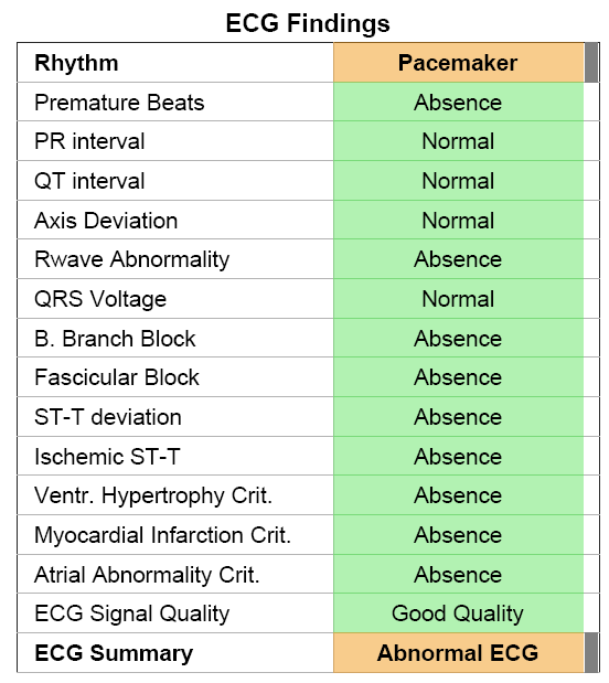
ECG
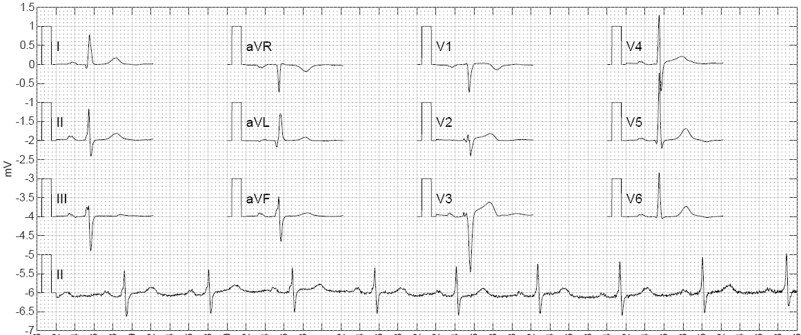
AI automated ECG interpretation detected pacemaker spikes; otherwise ECG signal is in the normal range.
PCG
PCG result shows an increased fourth sound (S4) amplitude, inaudible frequencies, rather late S4, occur after P-wave, right before the first sound. The reason for this type S4 is in question.
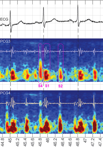
The increased amplitude of the second sound can be a sign, but alone it is not considered pathological.
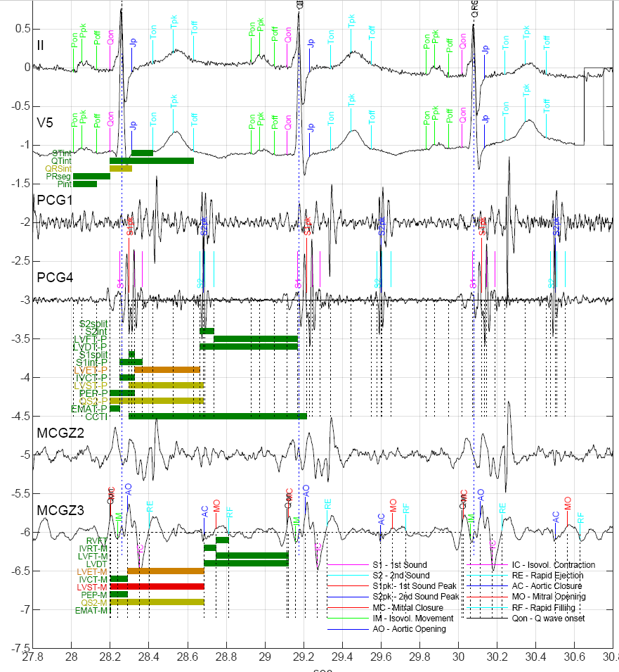
MCG
The MCG-based systolic time intervals show prolonged LV ejection time (LVET) or LV systolic time (LVST). However, the detection of Q onset seems to be shifted to earlier due to the pacemaker spikes, which may make uncertain the systolic time interval measurements.
MCG signal confirms the fourth sound activity, which means increased atrial systole. This is probably connected to diastolic dysfunction.
Echocardiographic Results
TTE confirms:
- preserved systolic function (LVEF=59% with Simpson Mod BP) which means slight improvement in LVEF;
- diastolic dysfunction type impaired relaxation – grade I (E/A=0.81, DT=213ms, E/e’(mean?)=9);
- pacemaker in the right chambers
Conclusion
A previous CHART report was not available for comparison purposes, but the current CHART report correctly indicates the echocardiographic results based on bio-signals available in primary care.
What is CHART?
Cardio-HART or CHART is an AI cardiac diagnostic system for the non-invasive detection of early onset of 93% of all common heart diseases, including heart failure and heart valve disease. It is a direct replacement for ECG, augmenting it with Echo functionality, that is indicated for use in clinical care settings, including primary care.
Noah RIchard TYler RaderMACher
|