
Myocardial infarction in question
November 1, 2022
Situation
A 54-year-old male patient has a previous (a month earlier) normal ECG, and the physician did not diagnose any problem.
CHART test is done currently, which shows a different picture.
CHART report interpretation
Risk Assessment
The blood pressure is high: 160/100. Otherwise no other cardiac-related risks.
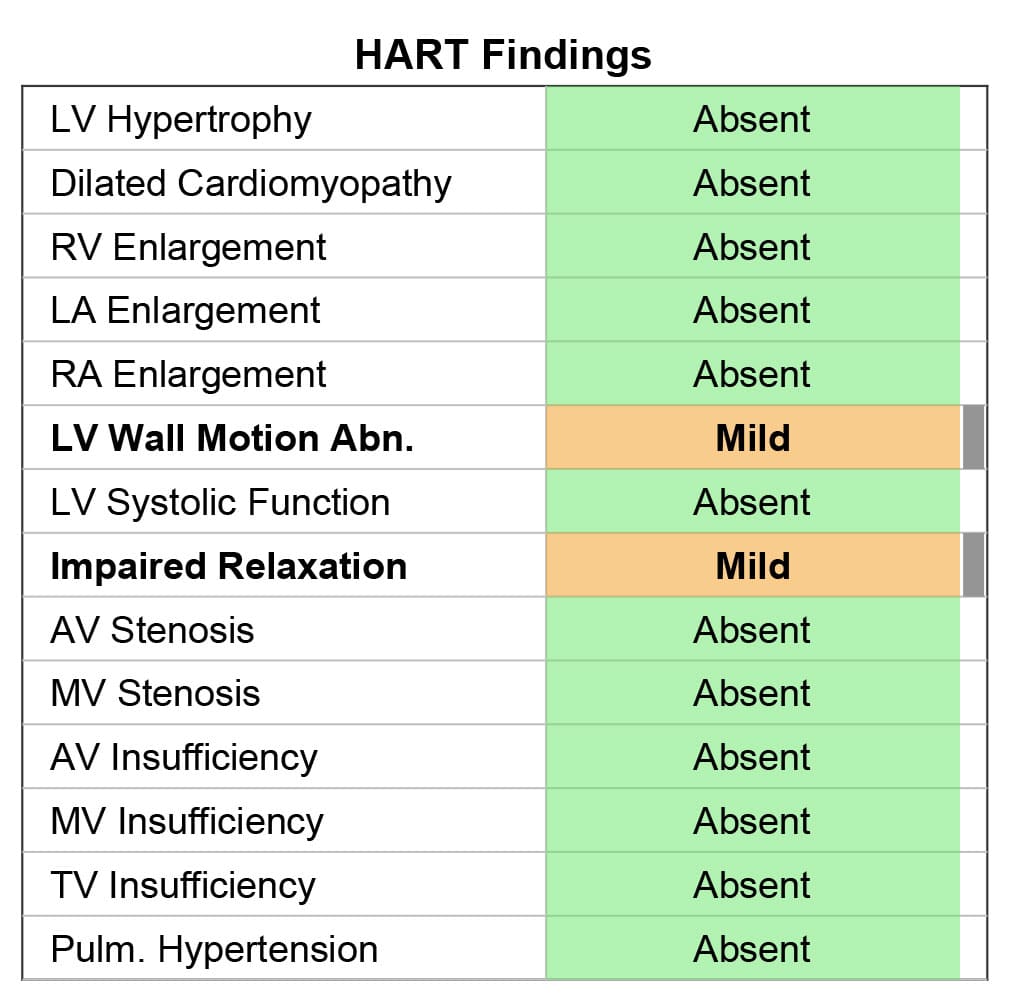
HART-findings and HF prediction
HART-findings show mild wall motion abnormality (WMA) and diastolic dysfunction type of impaired relaxation (DDIM). The WMA is probably connected to MI based on the ECG signal.
The DDIM is rather confirmed by the opening snap detected on the PCG signal and prolonged RVFT on MCG. The DDIM may also be connected to high diastolic pressure also.
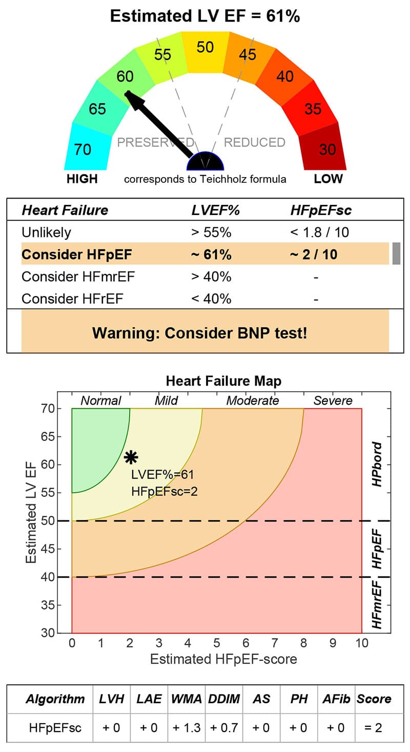
The estimated LVEF shows normal systolic function. The patient probably has no symptoms, but based on the HFpEF score, it fulfills the borderline criteria for a potential HFpEF patient.
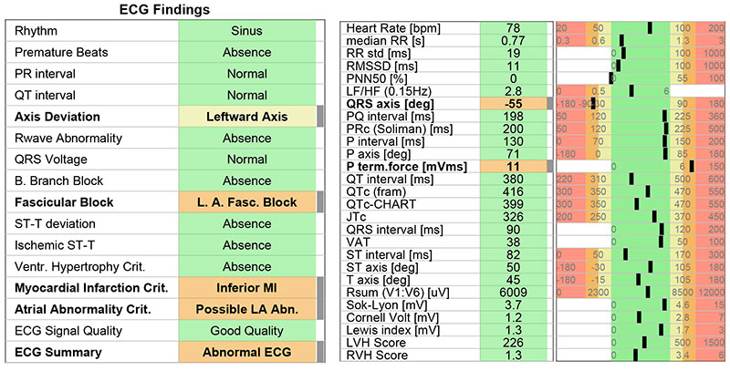
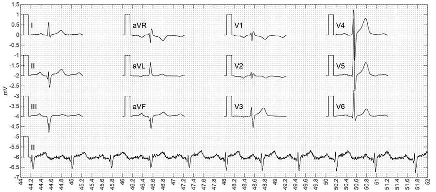
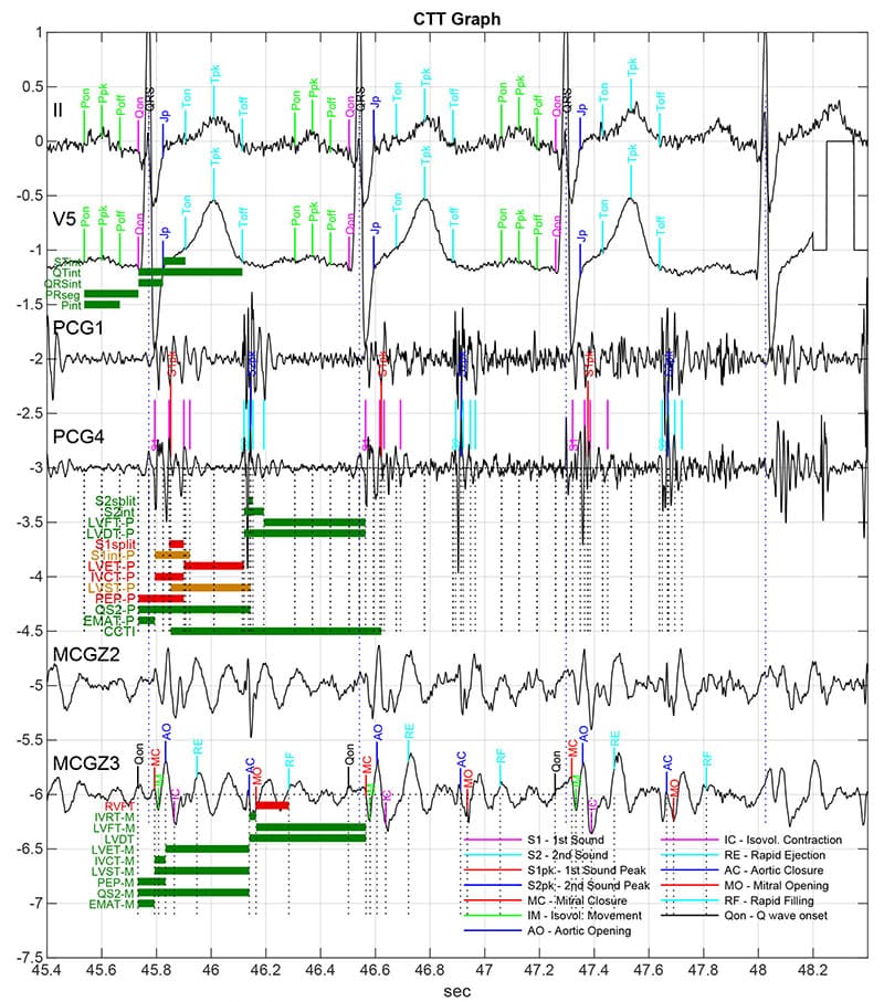
ECG
Leftward axis and left anterior fascicular block (LAFB) seem true positive.
Despite the ECG rule-based evidence, the inferior myocardial infarction is uncertain, because of the presence of LAFB. The ECG literature already discusses this problem[1].
The possible LA enlargement is detected based on P terminal force and longer PR interval (200), but LA enlargement was not confirmed by HART-findings. The literature reports high inconsistency between ECG P-mitral and ECHO-based LAE[2].
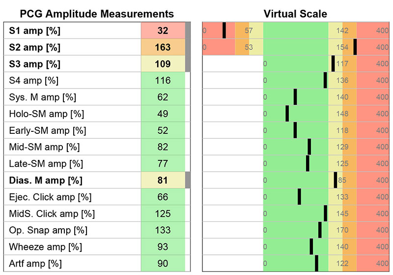
PCG
PCG signal shows wider but lower amplitude first sound and strong second heart sound. In addition, the length of the normal inactive systole (LVST) is shorter compared to the first sound periods, which is not typical.
MCG
The MCG signal shape is regular, the systolic time interval seems normal.
The pattern recognition of rapid ventricular filling time (RVFT) is less certain, but this may indicate any diastolic dysfunction.
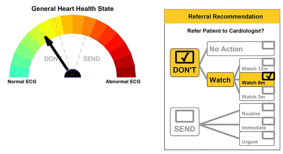
Conclusion
The patient has no symptoms, but the abnormalities detected by ECG and wall motion by HART-findings have a referral type of Watch 6 months for further analysis, as well as consider BNP test.
What is CHART?
Cardio-HART or CHART, is an AI cardiac diagnostic system for the non-invasive detection of the early onset of 93% of all common heart diseases, including heart failure and heart valve disease. It is a direct replacement for ECG, augmenting it with Echo functionality, that is indicated for use in clinical care settings, including primary care.
[1] Sohi, Gurbachan S., and Nancy C. Flowers. "Distinguishing features of left anterior fascicular block and inferior myocardial infarction as presented by body surface potential mapping." Circulation 60.6 (1979): 1354-1359.
Cooper, M. J., et al. "Diagnosis of inferior myocardial infarction in the presence of left anterior hemiblock." Australian and New Zealand Journal of Medicine 17.1 (1987): 47-50.
[2] Tsao, Connie W., et al. "Accuracy of electrocardiographic criteria for atrial enlargement: validation with cardiovascular magnetic resonance." Journal of Cardiovascular Magnetic Resonance 10.1 (2008): 1-7.